
Figure 1.
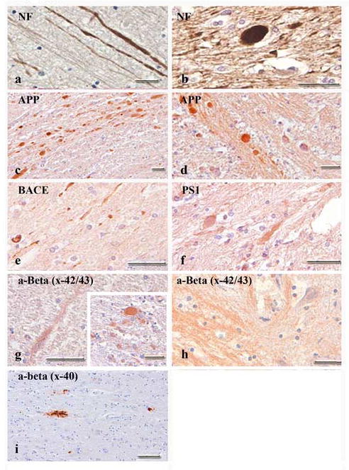
Bright-field photomicrographs showing pathological protein accumulation associated with axonal pathology in humans. Neurofilament and amyloid precursor proteins (APP) were both found in axonal swellings and bulbs following TBI (a-d). Enzymatic agents of APP cleavage (BACE and PS1) were also present, although to a lesser extent (e, f). Antibodies specific for Aβ revealed axonal swellings (g) and bulbs (g, inset) positive for Aβx-42/43 throughout the white matter of subjects and within the brainstem of several young cases (h). A limited number of swellings and bulbs stained positive for Aβx-40. Infrequent amyloid deposits in subjects over 70 years of age were seen with Aβx-40 (i). Scale bars = 50 μm.