
Figure 2.
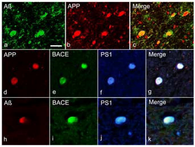
Multiple immunofluorescent staining showing co-accumulation of APP, BACE, PS1, and Aβ in axons following TBI. Double-labelling revealed co-accumulation of APP and Aβ1-42 in multiple axon bulbs (a-c). Further immunohistochemical staining showed co-accumulation of BACE and PS-1 with APP (d-g) and Aβ1-42 (h-k).Scale bars = 50 μm.