
Figure 3.
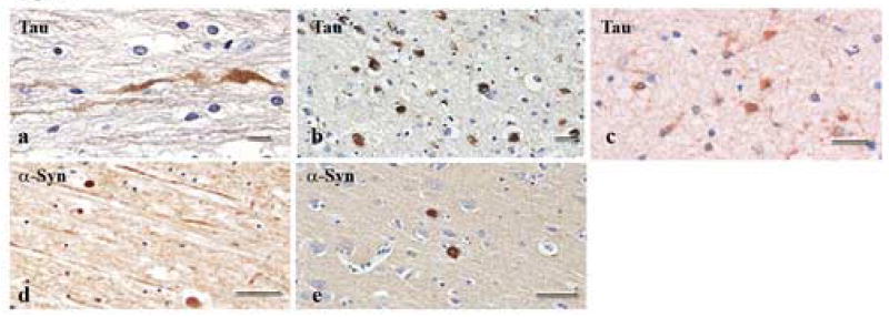
Bright-field photomicrographs showing accumulation of tau and α-syn proteins in axonal swellings and bulbs. Of the 18 TBI cases examined, only 2 stained for tau. Tau staining was observed in axonal swellings (a) and bulbs (b) following injury, and, to a lesser extent, in glial cells (c). α-syn was also present within axonal swellings (d) and bulbs (e) in a majority of the injured subjects. Scale bars = 50 um.