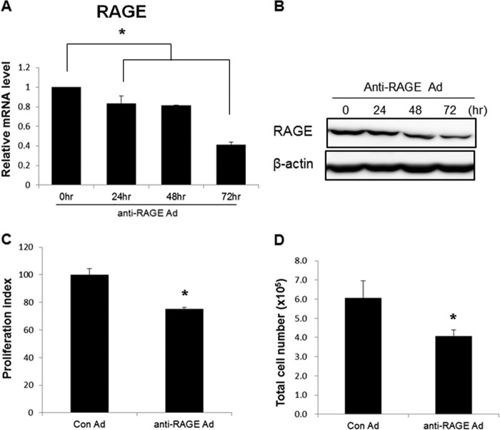
FIGURE 1.
Anti-RAGE adenovirus inhibited proliferation of WT 9-12 cells. A, reduction of the RAGE transcript in a time-dependent manner. Approximately 3.5 × 10⁵ cells were seeded in 100-mm dishes, and anti-RAGE adenovirus (m.o.i. = 200) was added. Fresh media were added after 24 h. The cells were harvested at the indicated time points. B, decrease in RAGE protein in a time-dependent manner. C, relative viability of infected cells was measured using the XTT assay (m.o.i. = 200, 72 h after infection). D, total number of cells in each group (m.o.i. = 200, 72 h after infection). Cells were stained with trypan blue and counted. Data are mean ± S.D. (error bars). *, p < 0.05.

