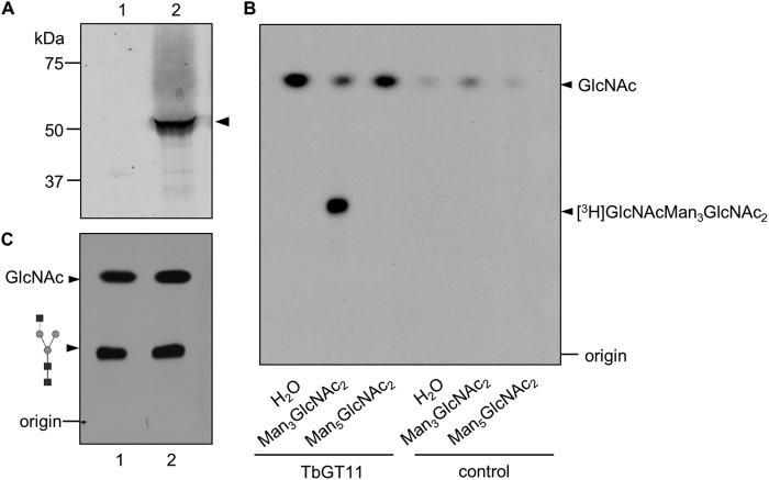
FIGURE 8.
TbGT11-HA3 expression and in vitro activity assay. A, Trypanosome bloodstream-form cell lysates from WT cells (lane 1) or cells transfected with pLEW82-GT11-HA3 (lane 2) were separated by SDS-PAGE and analyzed by Western blotting with a rabbit anti-HA antibody. B, TLC autofluorography of in vitro reaction products. After incubation of TbGT11-HA3 attached to anti-HA-conjugated magnetic beads with UDP-[3H]GlcNAc as well as the acceptor substrates Man3GlcNAc2, Man5GlcNAc2, or no acceptor (H2O), reaction products were separated by TLC (lanes 1–3). As a negative control, anti-HA magnetic beads incubated with lysates from cells not expressing TbGT11-HA3 were used (lane 4-6). C, the obtained [3H]GlcNAcMan3GlcNAc2 reaction product was separated by TLC before and after α1–2,3 mannosidase treatment and visualized by fluorography.

