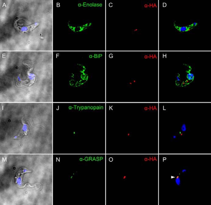
FIGURE 9.

Golgi localization of TbGT11. Fixed and permeabilized bloodstream-form parasites expressing TbGT11-HA3 were co-stained with anti-HA antibodies to detect TbGT11 localization (red) and either anti-Enolase (A–D), anti-BiP (E–H), anti-trypanopain (I–L), or anti-Golgi reassembly stacking protein (GRASP) (M–P) to detect the cytosol, ER, lysosome, or Golgi apparatus, respectively (green). Cells were counterstained with DAPI (blue) to reveal nuclei and kinetoplasts. Merged DAPI/DIC images are presented on the left and merged three-channel fluorescence images are presented on the right. Prominent co-localization is indicated by an arrowhead (P, yellow). No staining of untransfected (non-epitope-tagged) cells was detected under the same conditions with anti-HA antibodies (data not shown).
