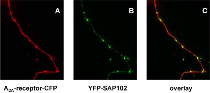
FIGURE 6.

Colocalization of A2A receptors and SAP102 in hippocampal neurons. Cultures of dissociated rat hippocampal neurons were transfected with plasmids coding for the CFP-tagged A2A receptor (A2A-receptor-CFP) and YFP-tagged SAP (YFP-SAP102). Images were captured in both the CFP (A, red pseudocolor) and the YFP channel (B, green pseudocolor), noise-corrected, and converted into an 8-bit RGB image. An overlay image (C, red, green, and yellow pseudocolors; scale bar = 19 μm) was generated in MATLAB by combining the two channels. Regions with both proteins appear in yellow pseudocolor. Data are representative of >10 neurons in three independent transfections.
