
FIGURE 6.
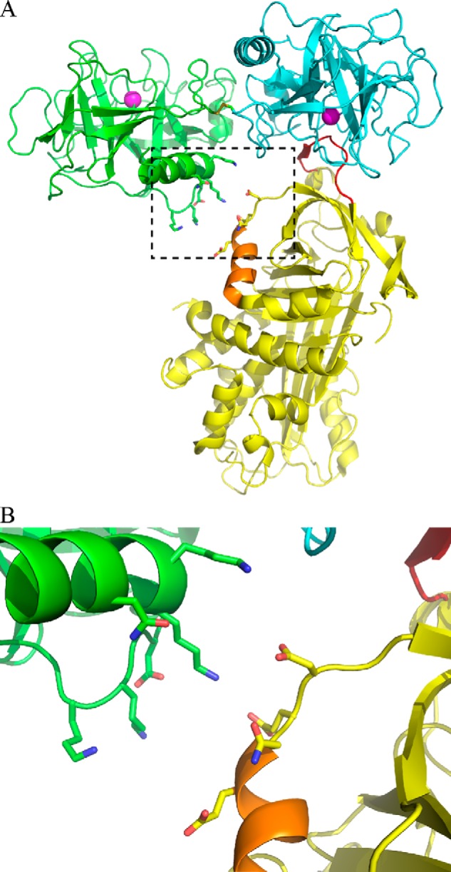
Model of the mouse granzyme A and Serpinb6b interaction. A, crystal structure of dimeric human GzmA (monomers are colored in green and cyan; PDB accession 1ORF) was aligned with the Michaelis complex (PDB accession 1OPH) between S195A trypsin (yellow) and M358R antitrypsin (aligned to the cyan GzmA monomer, not shown). Helix H is shown in orange. B, expanded view of the potential exosite interaction between mGzmA and Serpinb6b.