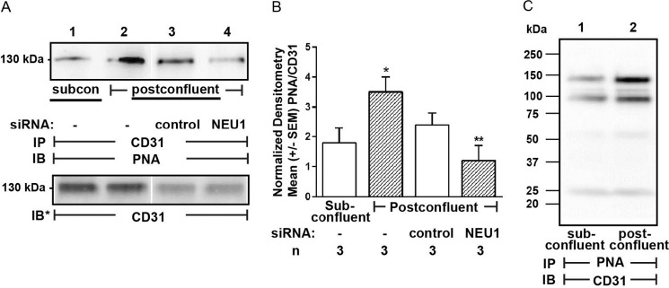
FIGURE 7.

NEU1 desialylates CD31 in postconfluent ECs. A, HPMECs (lanes 1 and 2) or HPMECs transfected with NEU1-targeting (lane 4) or control siRNAs (lane 3) were allowed to achieve subconfluent (lane 1) or postconfluent (lanes 2–4) states and lysed, and the lysates were immunoprecipitated with anti-CD31 antibody. The CD31 immunoprecipitates were processed for PNA lectin blotting. To control for protein loading and transfer, the blots were stripped and reprobed for CD31. B, densitometric analyses of the blots in A. Vertical bars, mean ± S.E. (error bars) PNA signal normalized to CD31 signal in the same lane on the same blot (n = 3). *, significantly increased PNA/CD31 densitometry of postconfluent versus subconfluent cells at p < 0.05. **, significantly decreased PNA/CD31 densitometry compared with control siRNA-transfected cells at p < 0.05. C, lysates from subconfluent (lane 1) and postconfluent (lane 2) HPMECs were incubated with PNA immobilized on Sepharose beads, and the PNA-binding proteins were processed for CD31 immunoblotting. IP, immunoprecipitation; IB, immunoblot; IB*, immunoblot after stripping. Molecular mass in kDa is indicated on the left. Each blot is representative of three independent experiments.
