
FIGURE 9.
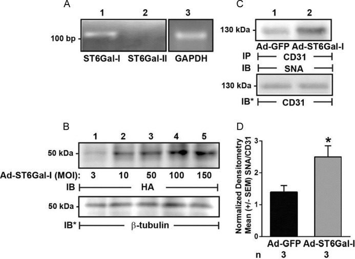
Increased ST6GAL-I expression in HPMECs increases α2,6-linked CD31 sialylation. A, total cellular RNA was extracted from HPMECs and processed for RT-PCR for ST6GAL-I and -II. GAPDH was run as an internal control. Base pairs (bp) are indicated on the left. B, HPMECs were infected with increasing MOIs of Ad encoding for HA-tagged ST6GAL-I, and after 48 h, they were lysed, and the lysates were processed for immunoblotting with anti-HA antibody. To control for protein loading and transfer, blots were stripped and reprobed for β-tubulin. C, HPMECs were infected with Ad-ST6GAL-I (lane 2) or Ad-GFP (lane 1), each at MOI = 200, and after 48 h, they were lysed, and the lysates were immunoprecipitated with anti-CD31 antibody. The CD31 immunoprecipitates were processed for SNA lectin blotting. To control for protein loading and transfer, blots were stripped and reprobed for CD31. For A–C, each blot is representative of three independent experiments. IP, immunoprecipitation; IB, immunoblot; IB*, immunoblot after stripping. Molecular mass in kDa is indicated on the left. D, densitometric analyses of blots in C. Vertical bars, mean ± S.E. (error bars) SNA signal normalized to CD31 signal in the same lane on the same blot (n = 3). *, significantly increased SNA/CD31 densitometry of Ad-ST6GAL-I-infected ECs compared with the Ad-GFP-infected control ECs at p < 0.05.