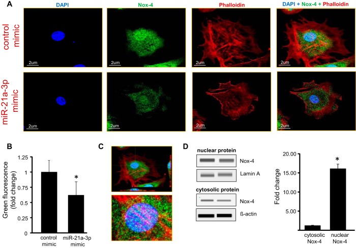
FIGURE 8.
Lowered Nox-4 in response to treatment with miR-21a-3p mimic. A, EOMA cells with or without miR-21a-3p mimic were labeled with fluorescent probes DAPI (blue) for nucleus, phalloidin (red) for actin and (green) for Nox-4. Confocal microscopy showed that Nox-4 was localized in perinuclear area in EOMA cells, and there was significant decrease in Nox-4 fluorescence intensity in miR-21a-3p delivered cells. B, fluorescence intensity of images was quantified using Olympus FV10-ASW software. C, Z-stack image showing Nox-4 localized mostly in the perinuclear space. D, immunoblot analyses of nuclear and cytosolic fractions of EOMA cell lysates compare relative distribution of Nox-4 protein. The results are expressed as means ± S.D. of at least three independent experiments. *, p < 0.05.

