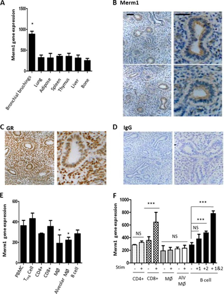
FIGURE 2.
Merm1 expression in normal human tissues. A, the expression of Merm1 was measured in a profile of normal human tissue samples acquired and analyzed by Affymetrix gene array. Sample preparation and analysis is described under “Experimental Procedures.” Independent samples were used: bronchial brushings = 7; lung = 100; spleen = 31; adipose = 113; thymus = 67; liver = 32; bone = 12. Analysis was carried out using a one-way ANOVA, with Tukey's post-hoc test (*, p < 0.05). B–D, Merm1 and GR expression in human fetal lung. Normal, human fetal lung explants were prepared, fixed, and analyzed by immunohistochemistry. Antibody binding was disclosed by 3,3-diaminobenzidine staining (brown), with nuclei counter-stained with toluidine blue. E, Merm1 gene expression in hematopoietic cells. Human Affymetrix gene expression databases (see “Experimental Procedures”) were interrogated for Merm1 expression. Independent samples were analyzed; n = 4, except alveolar macrophages, n = 5, and macrophages, n = 9. *, p < 0.05 one way ANOVA followed by Bonferroni test. F, Merm1 expression in stimulated hematopoietic cells. Human Affymetrix gene expression databases (“Experimental Procedures”) were interrogated for Merm1 expression. CD4 and CD8 lymphocytes were stimulated with anti CD3 (n = 4 for each group), macrophages (MØ) were stimulated with LPS (6 h treatment) (n = 9 for each group); alveolar macrophages (Alv MØ) were unstimulated (n = 5) and stimulated with LPS (6 h n = 15); B cells were stimulated with CD40 ligand (1), anti-B cell receptor antibody (2), or both (48 h treatment) (n = 4 for each group). ***, p < 0.001, one-way ANOVA, with Bonferroni post-hoc test; NS, not significant.

