FIGURE 7.
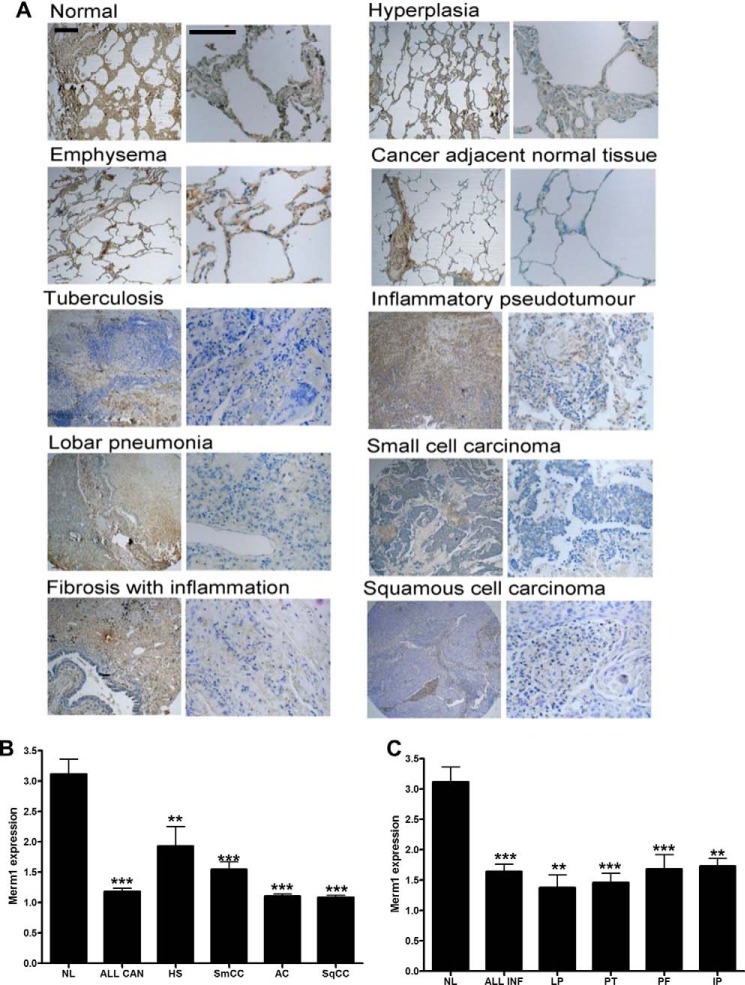
Merm1 expression in Lung diseases. A, Merm1 expression in a human lung disease tissue array. Human lung sections from Insight Biotechnology (LC487) were analyzed for Merm1 protein expression by immunohistochemistry; anti-Merm1 antibody (1:100). Low and high magnification views are presented for the different pathological states analyzed. The expression of Merm1 in each core sample was estimated by three masked observers scoring four microscope fields for each core. Scale bar, 100 um). Specific antibody binding was disclosed by 3,3-diaminobenzidine (brown) and nuclei counterstained with toluidine blue. B and C, the quantification of Merm1 expression seen in the different pathological states in (A) is presented. Expression was estimated on an arbitrary scale from 1–4, with 4 being very intense staining. Cancer pathology is presented in B, and inflammatory pathology is presented in C. NL, normal lung; All Can, all cancer states combined; HS, hyperplasia of the stroma; SqCC, squamous cell carcinoma; AC, adenocarcinoma; SmCC, Small cell carcinoma. C, normal lung repeated from B. All infect, all infection scores were combined; LP, lobar pneumonia; PT, pulmonary tuberculosis; PF, pulmonary fibrosis; IP, inflammatory pseudotumor. Analysis was by one-way ANOVA with Bonferroni post hoc tests. **, p < 0.01, ***, p < 0.001 compared with normal lung.

