FIGURE 1.
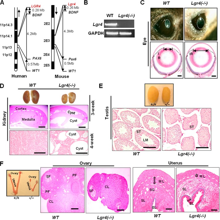
Lgr4 deletion leads to aniridia, polycystic kidney disease, and reproductive defects in mice. A, genetic loci of Lgr4, BDNF, Pax6, and WT1 in synteny chromosome fragments of human 11P and mouse 2E. Mb, megabases. B, RT-PCR results of Lgr4 expression in P0 wild type and Lgr4 depletion mouse tails. C, 1-month-old wild type and Lgr4−/− mouse eyes under strong light (top) demonstrated a significantly bigger pupil (white circle) in Lgr4−/− mouse eyes. H&E-stained samples of indicated mouse eyes showed a bigger pupil diameter in Lgr4−/− eyes (bottom). D, deletion of Lgr4 induces polycystic kidney disease. Cross-section of 3-week-old wild type and Lgr4−/− kidneys, with cysts in the Lgr4−/− kidney (top). The middle panel is a magnified H&E stained section of the top panel. H&E-stained 4-week-old wild type and Lgr4−/− kidney sections showed polycystic disease in the Lgr4−/− kidney (bottom). Representative photos are of n = 9 mice. E, Lgr4−/− mice present testicular development deficits. Representative 4-week-old wild type and Lgr4−/− testes are shown (top). H&E-stained sections (bottom) come from the top panel and show smaller seminiferous tubules and lumen formation defects in Lgr4−/− mice. F, representative 4-week-old wild type and Lgr4−/− ovary and H&E-stained samples are shown, with dramatically smaller size and fewer follicles in Lgr4−/− ovary. Lgr4−/− uterus had a significantly thinner muscle layer, almost no secretory glands, enhanced inner epithelial layer and abnormal stromal layer structure. IR, iris; LM, lumen; ST, seminiferous tubule; PF, primary follicle; SF, secondary follicle; CL, corpus luteum; ML, muscle layer; SG, secretory gland; SL, stroma layer; EL, inner epithelial layer. Scale bar = 20 μm.

