FIGURE 2.

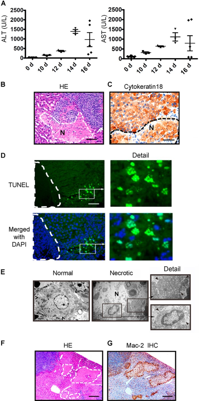
Energy deprivation in the tumor-bearing liver is positively associated with liver injury and hepatocyte necrosis in colon cancer liver metastasis. A, the serum ALT and AST activity mice were determined by immune complex kinase assays at the time indicated in both mice injected with SL4 and mice not injected with SL4. The injury to the liver worsened as the tumor developed in the liver (n = 8). B, the livers were harvested 12 days after SL4 injection, paraffin-sectioned, and subjected to hematoxylin and eosin staining. The dashed line and letter N indicate the necrotic mass with weak staining of cell nuclei. Scale bar, 100 μm. C, the livers were frozen-sectioned and subjected to immunohistochemistry with a specific antibody against cytokeratin 18 that recognized hepatocytes. The dashed line and letter N indicate the necrotic cells of hepatocytes with weak staining of cell nuclei. Scale bar, 100 μm. D, a tumor-bearing liver that was paraffin-sectioned on day 12 was subjected to TUNEL staining to indicate the dead cells. The dashed line indicates the tumor region. Scale bar, 100 μm. E, electron microscopy images show necrotic hepatocytes in the tumor-bearing liver on day 12. The arrowhead indicates the cell border. Scale bar, 100 μm. F and G, hematoxylin and eosin (HE) and IHC staining with Mac-2 antibody show the necrotic masses and the accumulation of macrophages around the masses in the tumor-bearing liver on day 12. The dashed line indicates the necrotic mass. Scale bar, 200 μm. d, day(s).
