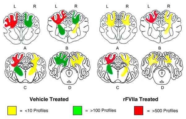
Fig.7.
Schematic representation of the distribution and severity of axonal pathology in various coronal planes of the brain at 3 days post-injury. In all animals the contusion injury was performed on the left side. (A) septal nuclei and anterior commissure level, (B) rostral-thalamus level, (C) caudal–hippocampal level, (D) occipital cortical brain stem level.

