
Figure 5.
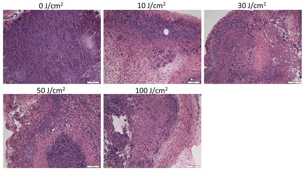
Histological pictures of A431 tumors, which were treated with PIT at 0, 10, 30, 50 and 100J/cm2 sections are shown. All specimens were stained with hematoxylin and eosin. Microscopic evaluation of PIT-treated tumors revealed diffuse necrosis and micro-hemorrhage with scattered clusters of damaged tumors cells after higher exposure of NIR light, 50 and 100J/cm2. At the highest light exposure group, 100J/cm2, necrotic damage was more intense and fewer tumor cells remained. In contrast, when only 10J/cm2 of NIR light exposed, necrotic cell damage was found in only limited areas within the tumor while substantial amounts of healthy cancer foci remained. Cancer cell damages in tumor after varying doses of NIR light exposure correlated well to the kinetic changes which were detected by dynamic fluorescence imaging with ICG. Scale bars represent 100μm.