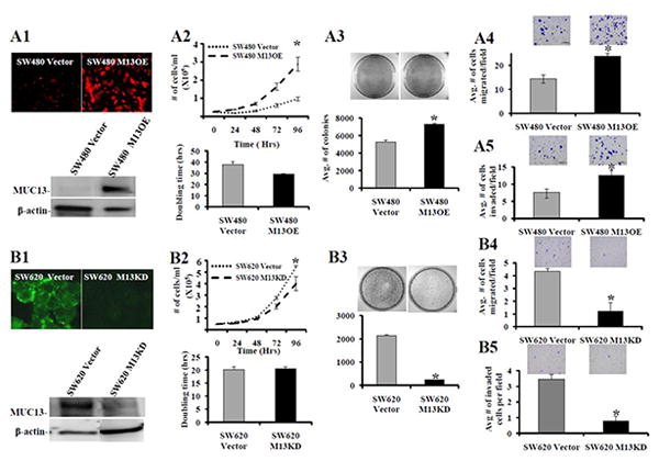
Fig. 1. MUC13 expression enhances the tumorigenic features of colon cancer cells.

(A) SW480 cells were transfected with GFP tagged full length MUC13 to obtain MUC13 over-expressing (SW480 M13OE) cells. Vector control (SW480 Vector) cells were also selected. (B) SW620 cells were transduced with MUC13 specific shRNA lentiviral particles to obtain MUC13 knock-down (SW620 M13KD) cells. Vector control (SW620 Vector) cells were also obtained. The selected cell populations were screened for MUC13 by immunofluorescence (A1 and B1, top) and confirmed by Western blot (A1 and B1, bottom). Cell growth (A2, B2, top), cell doubling time (A2 and B2, bottom), colony formation (A3 and B3, top and bottom), cell migration (A4 and B4), and cell invasion (A5 and B5) assays were performed with MUC13 over-expressing and MUC13 knock-down cells, respectively, as described in experimental procedures. Representative images are shown above bar graph for the corresponding assays. Bar indicates the mean, error bar indicates the SEM, N=3, * P<0.05.
