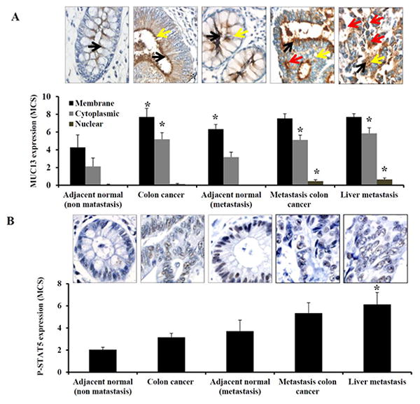
Fig. 5. P-STAT5 expression is associated with MUC13 expression in colon cancer tissues.

Tissue microarrays containing tissues from patients with non-metastatic disease (adjacent normal and colon cancer), and from patients with metastatic disease (adjacent normal, metastatic colon cancer, and liver metastasis tissues) were processed for immunostaining using MUC13 and P-STAT5 antibodies. Representative images of (A) MUC13 and (B) P-STAT5 immunostaining are shown and quantification was done as described in experimental methods. Black, yellow and red arrow indicates membranous, cytoplasmic and nuclear MUC13 expression respectively. Bar indicates the mean, error bar indicates the SEM, * P<0.05.
