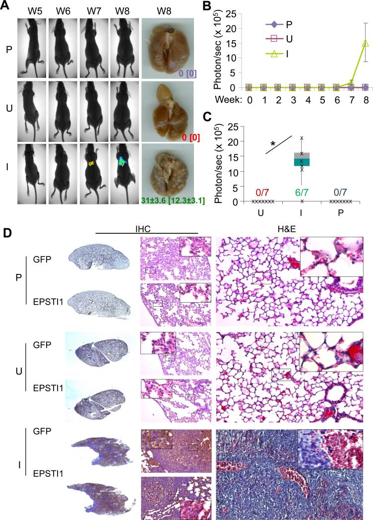
Figure 4.
EPSTI1 promotes the lung metastasis of breast cancer. The MCF7-iE1 cells or the parental cells (P) were injected into the tail vein. The mice injected with MCF7-iEPSTI1 cells were fed with the Dox Diet (I, EPSTI1 induction) or Control Diet (U, no EPSTI1 induction). The mice injected with the parental cells were feed with normal food. The lung metastasis was followed up for 8 weeks by BLI or stereomicroscopy at week 8 as described in the Materials and Methods. (a) Representative images Representative images with average number and standard deviation of metastatic tumor nodules per mouse visible on the surface of the lungs or stained positive for GFP in the tissue section shown in the brackets. (b) Quantitative results of the volumes of the metastasis. (c) Metastatic incidence at week 8. *P < 0.05. (d) H&E and IHC staining with GFP or EPSTI1 antibody confirming the human origin of the lung metastases and the effective induction of EPSTI1 expression in the tumors.

