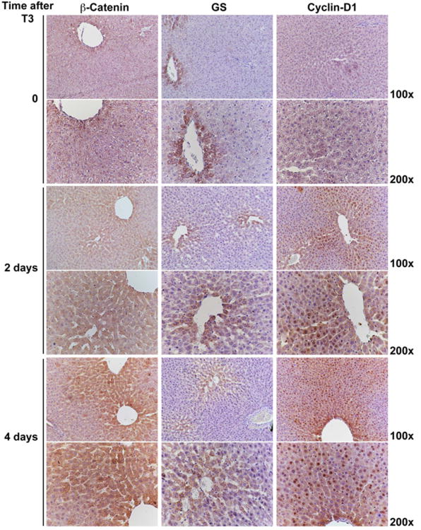
Figure 1. Activation of β-catenin signaling in rat livers after T3-feeding.
Immunostaining shows β-catenin localizing to the hepatocyte membrane in the control livers, while it accumulates in the hepatocyte cytoplasm in T3-fed rats at 2 days. A progressive shift of β-catenin stabilization from zone I towards zone II along with nuclear translocation of β-catenin-positive is observed at day 4 of T3 feeding. A concomitant increase in the number of GS-positive cells around central vein is observed at 4 days after T3. Progressive Cyclin-D1 nuclear staining is evident in zone I at 2 days, and in zone I and II at 4 days of T3 feeding as compared to the control livers. Four rats per group were used for this study.

