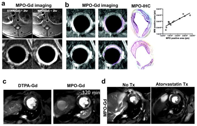
Fig. 6.
(a) Delayed contrast MRI at two hour shows brighter signal in MPO-Gd imaging as compared to DTPA-Gd in rabbit model of atherosclerosis (reproduced from Ronald et al. [84] with permission). (b) MPO-Gd also precisely localizes focal areas of inflamed plaque as outlined and confirmed with MPO immunohistochemistry visually as well as correlation analysis. (c) At two hours post contrast injection, much of the DTPA-Gd is washed away however MPO-Gd still lights up the infarcted myocardium in mouse model. (d) Follow up of atorvastatin treatment response with MPO-Gd imaging reveals decreased inflammation and in vivo MPO activity (modified from Nahrendorf et al. [85] with permission).

