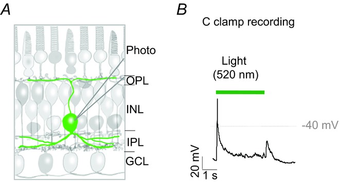
Figure 1.

A, schematic drawing illustrates the location of the interplexiform cell and its extended processes. B, current clamp recording shows the voltage responses of an interplexiform cell to the onset and offset of a 3 s green (520 nm) stimulus. GCL, ganglion cell layer; INL, inner nuclear layer; IPL, inner plexiform layer; OPL, outer plexiform layer; Photo, photoreceptor.
