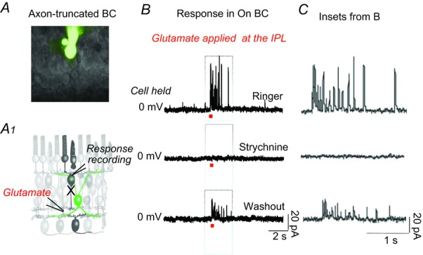
Figure 2.

A, axon-truncated ON BC in the dark-adapted retinal slice preparation following Lucifer Yellow staining. The experimental procedure is depicted in the schematic drawing below (A1). B, synaptic currents recorded from an axon-truncated cell held at 0 mV in response to focal application of 1 mm glutamate at the IPL. The initial currents were completely suppressed by strychnine, and partially recovered after washout. C, inset boxes from (B) in an extended time scale. BC, bipolar cell; IPL, inner plexiform layer.
