Figure 1.

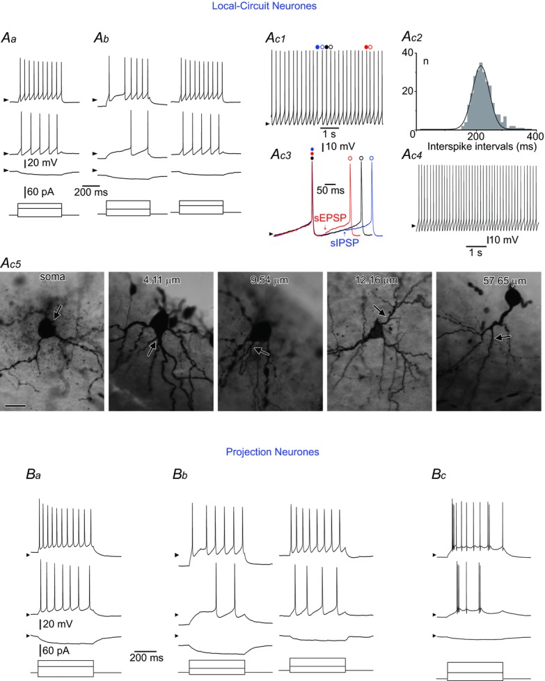
Aa, a tonic LCN. Ab, a gap firing LCN. The stimulation protocol was applied to the neurone kept at its resting membrane potential (RMP) (left) and after sustained depolarization to −70 mV (right); note the change of firing pattern to tonic. Ac1–4, a rhythmic LCN. Ac1, intrinsic firing at zero current injection. Spikes indicated by coloured circles are shown superimposed in Ac3. Ac2, distribution of interspike intervals for the neurone shown in Ac1 (bin width: 10 ms; 259 intervals analysed). Data are fitted with a Gaussian function (mean: 220 ms; σ, 29 ms). Ac3, interspike intervals: control (black), shortened by a spontaneous EPSP (sEPSP, red) and prolonged by a spontaneous IPSP (sIPSP, blue). Ac4, rhythmic firing in the presence of the mixture of 10 μm CNQX, 100 μm picrotoxin and 1 μm strychnine (in the neurone in Ac1). All spontaneous EPSPs and IPSPs were blocked. Ac5, somatic and dendritic origins of the axon initial segment in five different LCNs showing distances between the axon origin point and the soma. Scale bar: 25 μm. Ba, a tonic PN. Bb, a gap firing PN. The stimulation was applied to the neurone kept at its RMP (left) and after sustained depolarization to −70 mV (right). Bc, a burst firing PN. Stimulation protocols are shown below current-clamp recordings; arrowheads indicate a potential of −70 mV.
