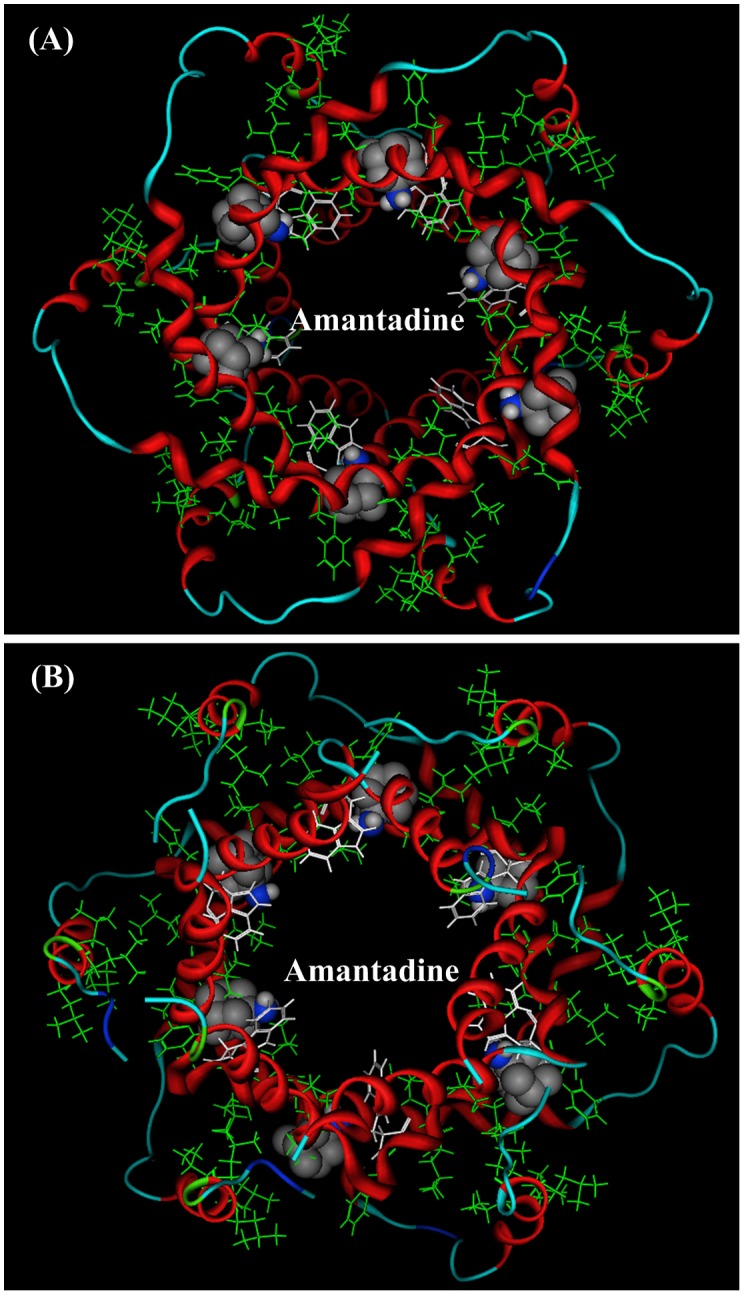
Figure 1. NMR solution structure (PDB code: 2M6X) of HCV p7 ion channel and the positions of ligand amantadine.

In p7 ion channel there are six equivalent hydrophobic pockets between the peripheral and pore-forming helices. The ligand amantadine (or rimantadine) is located in the hydrophobic cavities. The pocket consists of Phe 20, Val 25, Val26, Leu52, Val53, Leu55, and Leu56,. The amino group of amantadine on average points to the channel lumen [16]. (A) A view from the top of channel. (B) A view from the bottom of channel.
