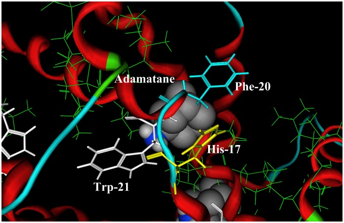
Figure 2. A close view of the binding pocket of amantadine in the p7 ion channel.
The hydrophobic residues (Phe20, Val25, Val26, Leu52, Val53, Leu55, and Leu56) are shown in green line drawing, which comprise the hydrophobic binding pocket of amantadine. The positions of three possible binding sites His17, Phe20, and Trp21 for the protonated pharmocophore group (−NH3 +) of amantadine are shown in yellow, light blue, and white, respectively. All three aromatic residues (His17, Phe20, and Trp21) are on the chain 2.

