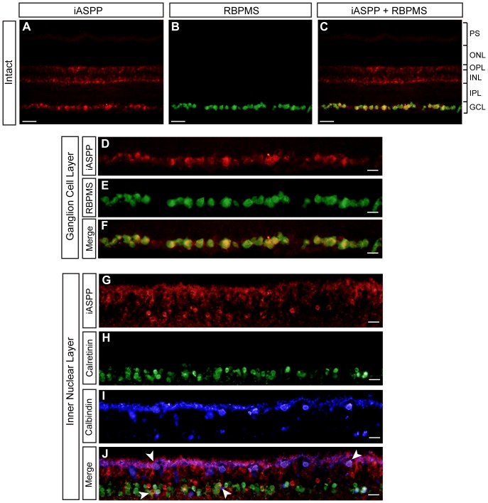
Figure 1. Adult RGCs express iASPP.
Endogenous retinal iASPP was detected by immunofluorescence in the ganglion cell layer (GCL) and inner nuclear layer (INL) (A, C, D, and G). iASPP staining in RGCs was confirmed using the RGC-specific marker RBPMS (D–F). iASPP was also detected in amacrine and horizontal cells, visualized with calretinin (H,J, arrows) and calbindin (I,J, arrows), respectively. Scale bars: (A–C) = 50 μm; (D–J) = 20 μm. PS: Photoreceptor Segments; ONL: Outer Nuclear Layer; OPL: Outer Plexiform Layer; INL: Inner Nuclear Layer; IPL: Inner Plexiform Layer; GCL: Ganglion Cell Layer.

