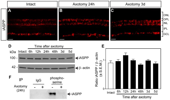
Figure 2. iASPP protein and phosphoserine levels after axotomy.
Retinal iASPP expression and localization did not change at 24 hrs or 3 days after optic nerve injury compared to intact eyes (A–C). Scale bar: 10 μm. Analysis of protein homogenates from axotomized retinas collected at 6, 12, 24, 48 hrs, 3 and 5 days confirmed that iASPP levels were similar to those in intact, non-injured retinas. The lower panel represents the same blot as in the upper panel but probed with an antibody that recognizes β-actin used to confirm equal protein loading (D). Densitometric analysis of western blots, showing the ratio of iASPP protein relative to β-actin, confirmed that there is no significant change in protein levels after injury (E) (ANOVA, p>0.05). Phosphoserine immunoprecipitation (IP) of intact and axotomized retinas probed with iASPP antibody revealed a decrease in phosphoserine iASPP at 24 hrs after axotomy. IP of retinal homogenates with an IgG antibody was included as control for non-specific interactions (F). ONL: Outer Nuclear Layer; OPL: Outer Plexiform Layer; INL: Inner Nuclear Layer; IPL: Inner Plexiform Layer; GCL: Ganglion Cell Layer.

