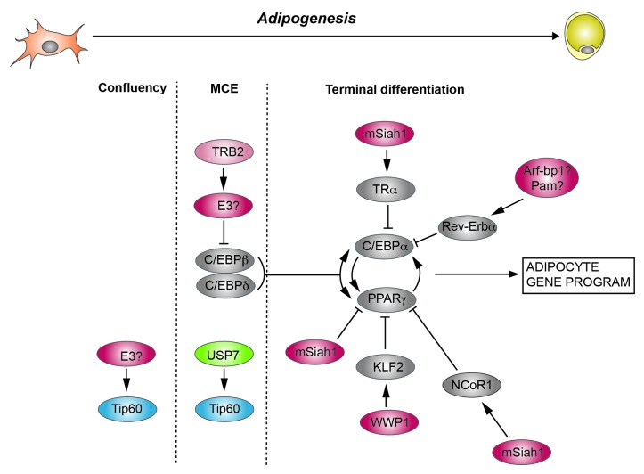
Figure 1. Protein (de)ubiquitination in adipogenesis. Indicated are the 3 stages of 3T3-L1 differentiation: growing to confluency, mitotic clonal expansion (MCE), and terminal differentiation. Also depicted are selected transcription factors and co-regulators that are subject to (de)ubiquitination. Ubiquitin E3 ligases are depicted in red, deubiquitinases in green. Please note that TRB2 itself is not an E3 ligase, but stimulates C/EBPβ ubiquitination through an unidentified E3 ligase.

An official website of the United States government
Here's how you know
Official websites use .gov
A
.gov website belongs to an official
government organization in the United States.
Secure .gov websites use HTTPS
A lock (
) or https:// means you've safely
connected to the .gov website. Share sensitive
information only on official, secure websites.
