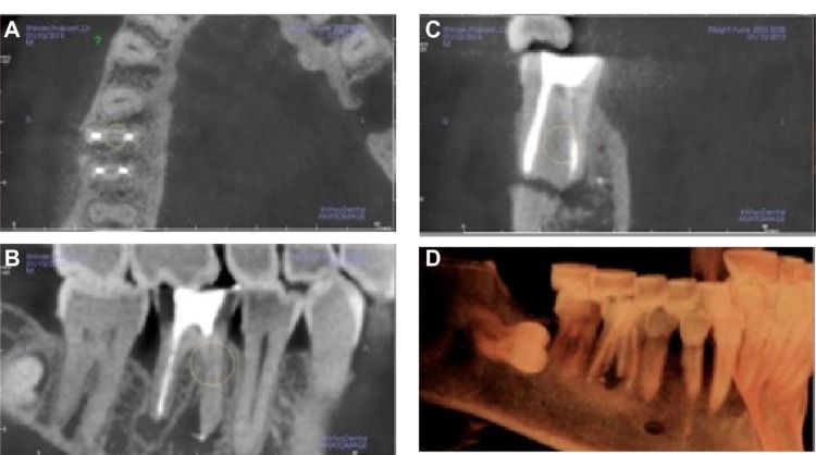
Figure 6.
CBCT image showing the missed canal in the mandibular molar.
Notes: Images courtesy of Dr Niranjan Vatkar, endodontist, Pune, India. (A) Missed canal seen on axial view. (B) Sagittal view showing large radiolucent lesion with mandibular first molar. (C) Cross sectional view showing missed canal. (D) 3D reconstruction showing osteolytic lesion with mandibular first molar.
Abbreviation: CBCT, cone-beam computed tomography.

