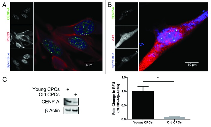
Figure 2. CENP-A declines in old CPCs. (A) Immunocytochemical analysis of CENP-A (green), the actin stain ph633 (red), and nuclear stain sytox (blue) in CPCs cultured in vitro. (B) Immunocytochemical analysis confirming CENP-A staining (green) co-localizes with c-kit (red). (C) Immunoblot demonstrating that CENP-A is significantly diminished in CPCs derived from old mice. Quantification shown in right panel (n = 3). *P < 0.05.

An official website of the United States government
Here's how you know
Official websites use .gov
A
.gov website belongs to an official
government organization in the United States.
Secure .gov websites use HTTPS
A lock (
) or https:// means you've safely
connected to the .gov website. Share sensitive
information only on official, secure websites.
