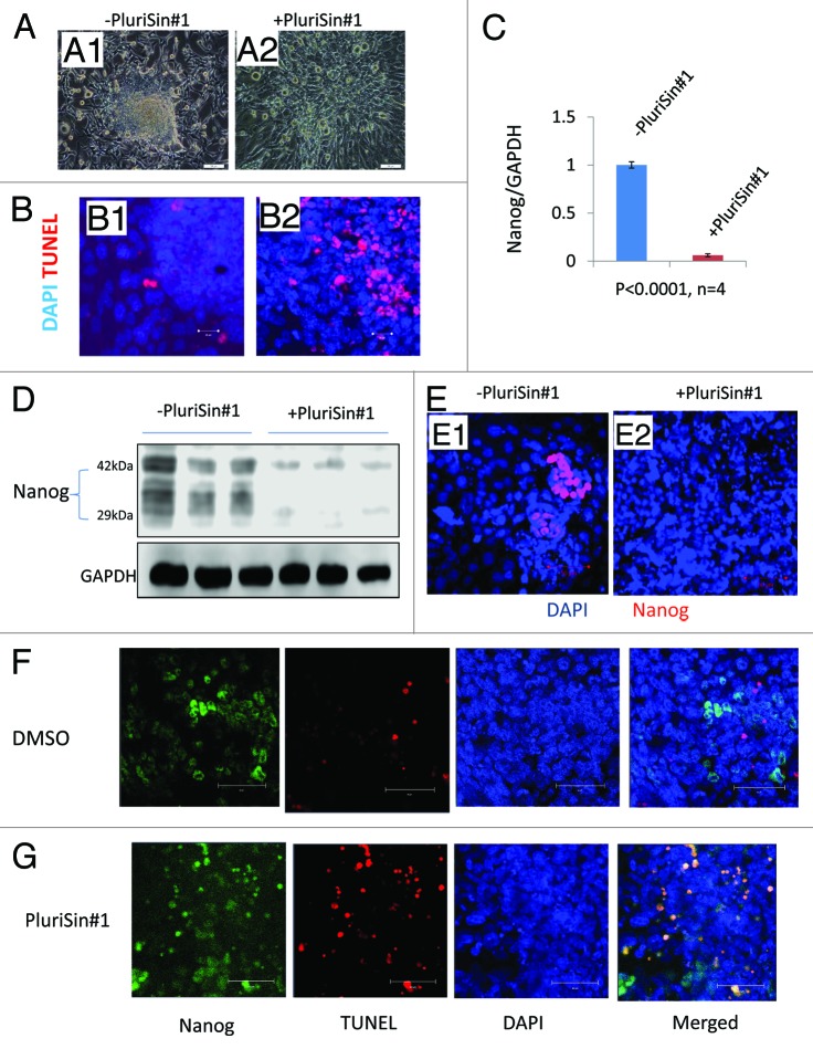
Figure 3. Effects of PluriSin#1 on Nanog-positive iPSD. (A1 and A2) Mouse iPSD were incubated with DMSO or PluriSin#1 for 4 d; note the appearance of cell death in the central region of iPSD treated with PluriSin#1; (B1 and B2) DMSO- and PluriSin#1-treated iPSD were assayed for apoptosis using TUNEL staining; note extensive TUNEL positivity in the center of PluriSin#1-treated iPSD; (C) Real-time RT-PCR analysis demonstrating the effect of PluriSin#1 on Nanog mRNA expression; (D) Protein was isolated from DMSO or PluriSin#1 treated IPSD and then used for immunoblotting; (E1 and E2) Immunostaining for Nanog demonstrating elimination of Nanog-positive iPSD cells by PluriSin#1. Nuclei were counterstained with DAPI; (F and G) Mouse iPSD were incubated with DMSO or PluriSin#1 for 1 d, a combined TUNEL and Nanog immunofluorescent staining demonstrating apoptosis of Nanog-positive iPSD cells by PluriSin#1. Nuclei were counterstained with DAPI.

An official website of the United States government
Here's how you know
Official websites use .gov
A
.gov website belongs to an official
government organization in the United States.
Secure .gov websites use HTTPS
A lock (
) or https:// means you've safely
connected to the .gov website. Share sensitive
information only on official, secure websites.
