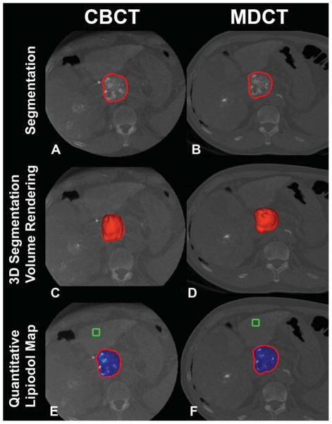
Figure 2.
3D volumetric semi-automatic evaluation of diffuse lipiodol retention in HCC on a representative case. Segmentation of the tumor (red circle) on CBCT at corresponding slice level as MDCT (A, B). 3D segmentation volume rendering on the same slice (C, D). Quantitative lipiodol color map of CBCT and MDCT (E, F). The box represents the location of the background ROI. The tumor volume on CBCT and on MDCT was 50.89cm3 and 50.78 cm3, respectively. The volume of lipiodol on CBCT and on MDCT was 42.87cm3 and 45.30 cm3, respectively.

