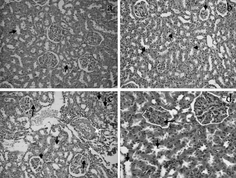
Fig. 2.
a. Image of mild dilatation (arrows) in the renal tubules of the control group (H&E, × 200). b. Image of mild tubular dilation (arrow) and tubular degeneration (small arrowheads) together with glomerular sclerosis (arrowhead) in renal tissue of the carnitine group (H&E, × 200). c. Image of focal glomerular necrosis (long arrows), degeneration and dilatation in the tubular epithelium (small arrows), expansion of Bowman’s capsule (arrowhead), inflammation (asterisks) and thickening of the blood vessel wall (white arrow) in renal tissue of the CM group (H&E, × 200). CM, contrast medium. d. Image of congestion (long arrow) and mild dilatation in the tubular epithelium (small arrows) in renal tissue of the CM+carnitine group (H&E, × 400). CM, contrast medium.

