
Figure 5. Olig1 regulates interneuron versus oligodendrocyte cell fate in neural stem cell cultures.
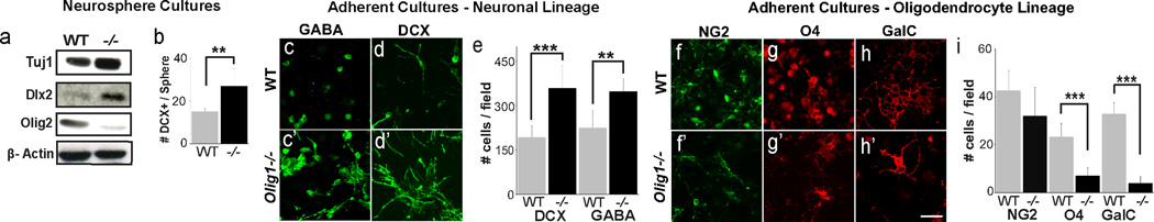
(a) Western blots from WT and Olig1−/− neurospheres for the neuronal protein Tuj1, Dlx2, and Olig2 showing increased expression of neuronal proteins and decreased expression of Olig2. (b) Quantification of number of DCX+ cells per neurosphere identified by immunohistochemistry (c–d) Representative images of neural progenitor monoloyer cultures derived from P3 WT and Olig1−/− SVZ, differentiated for 1 week and stained for DCX (c) and GABA (d). (e) Quantification of the number of DCX and GABA cells captured at 3 defined coordinates in chamber slide wells reveals increased numbers of DCX and GABA+ cells in Olig1−/− versus wild type. (f–h) Representative images of neural progenitor monolayer cultures derived from P3 WT and Olig1−/− SVZ, differentiated for 1 week and stained for NG2 (g), O4 (h) and GalC. (i) Quantification of the number of NG2, O4 and GalC cells captured at 3 defined coordinates in chamber slide wells reveals decreased numbers of O4 and GalC+ cells in Olig1−/− versus wild type. (For all quantifications: mean +/− SEM, n = 3 experiments, 4 slide wells per experiment; *p<.05, **p<.01, ***p<.005, 2 tailed unpaired student’s t test). (h) scale bar = 50 µm. See also Figure S5.