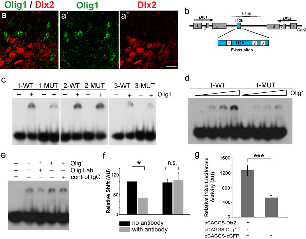
Figure 6. Olig1 is a direct repressor of Dlx1/2 at the I12B intergenic enhancer.
(a) 1 µm confocal projection demonstrating that Olig1 (green, a’) and Dlx2 (red, a“) does not colocalize Olig1 in VZ. (b) Schematic of the Dlx1/2 bigenic region showing location of I12B intergenic enhancer and 3 E-box sites. (c) Images of gels from electrophoretic mobility shift assays (EMSA) for the 3 I12B E-boxes (WT, wildtype and MUT, mutated) in presence or absence of Olig1 protein. Note the strongest and most specific affinity for E-box site 1. (d) Increasing concentrations of Olig1 protein show dose-dependent affinity of Olig1 for E-box 1 WT, but not for E-box 1 MUT. (e) Supershift assay demonstrates that Olig1 antibody, but not control IgG antibody inhibits binding of Olig1 protein to E-box site 1. (f) Quantification by densiometry of inhibition of DNA binding by incubation of Olig1 protein with Olig1 or control antibody (student T test * p <0.05) (g) Luciferase assays demonstrate that Olig1 is a transcriptional repressor capable of reducing Dlx2 induced I12B luciferase activity to 40% control levels (student’s t test ***p <0.001). (a”) scale bar = 50 µm. See also Figures S5 and S6.

