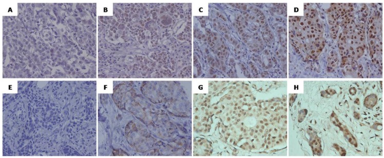
Figure 1.
Representative immunohistochemical staining of SRC-1(A-D) and Twist1 (E-H) in human breast cancer. (A, E) Negative staining, score 0. (B,F)Weak positive staining, (B) Score 6 (intensity 1, percentage 5), (F) Score 4 (intensity 1, percentage 3); (C, G) Moderate positive staining, (C) Score 7 (intensity 2, percentage 5),(G) Score 7 (intensity 2, percentage 5); ( D, H) Strong positive staining.(D) Score 8 (intensity 3, percentage 5), (H) Score 8 (intensity 3, percentage 5). Original magnification, ×400.

