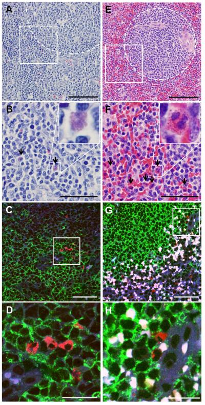
Fig 3.

Eos can be found in close proximity to B cell follicles in human tonsils and spleens. Human tonsils (A-D) and spleens (E-H) were stained with H&E (A-B, E-F) or using immunofluorescence (C-D, G-H) for specific visualization of Eos and B cells. In H&E stained images, B cell follicles are outlined in the dotted regions (A and E) and Eos are highlighted by black arrows (B and F). In immunofluorescence stained slides, anti-MBP labeled Eos in red, anti-CD19 labeled B cells in green. Autofluorescent red blood cells are white in these overlaid images. B, D, F, and H show higher magnifications of areas enclosed in white box in A, D, E, and F, respectively. Scale bar, 100 μm (A and E); 50 μm (B-C and F-G); 20 μm (D and H).
