Fig 6.

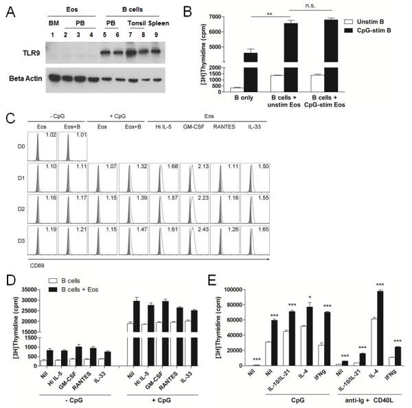
Activation of Eos is not required for the support of B cell proliferation. A, Eos isolated from human BM and PB and B cells isolated from PB, tonsils, and spleens were analyzed for TLR9 protein expression. Beta actin was used as loading control. B, B cells and Eos were stimulated independently with CpG. Proliferation of unstimulated and CpG-stimulated B cells was assessed either alone or in the presence of unstimulated or CpG-stimulated Eos. C, Eos were cultured alone or with B cells ± CpG or in the presence of various Eos-activating cytokines. Eos were analyzed for surface expression of CD69 on D0, D1, D2, and D3 by flow cytometry (solid grey ( ), isotype; open black (□), anti-CD69 mAb). Numbers in FACS plots represent △MFI. Data are representative of 3 independent experiments. D-E, B cells or B+Eos cultures were stimulated with various cytokines for Eos activation (D) or B cell activation (E) and proliferation was evaluated. * p<0.05; ** p<0.01; *** p<0.001; n.s., not significant.
