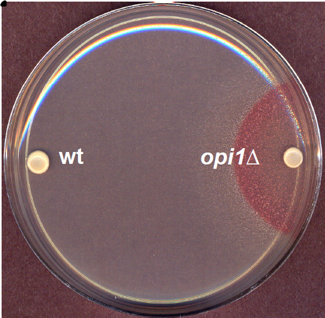
Figure 2. Overproduction of inositol (Opi−) phenotype of opi1Δ strain.
Wild type (wt) and opi1Δ cells were spotted on plates containing I− medium and incubated for 2 days at 30°C. A cell suspension of AID indicator strain, which grows only in the presence of inositol, was sprayed on the plates and incubated for a further 2 days at 30°C. Strains excreting inositol are visible as red halos around the strain being tested.

