Fig 5.


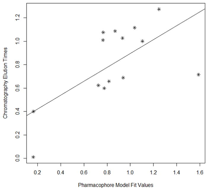
Fig 5A Scatter plot of chromatographic elution times and pharmacophore model (see Table 5). The best fit line for the data is also provided.
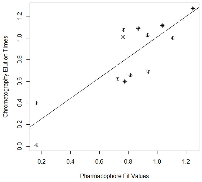
Fig 5B. Scatter plot of training set (Fig 6A) without Apigenin vs the experimental elution times. The best fit line for the data is also provided. (Pearson (r) : 0.8456, Spearman(r) = 0.7088
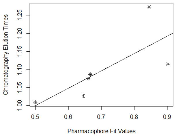
Fig 5C. Scatter plot of chromatographic elution times and pharmacophore model (see Table 6). The best fit line for the data is also provided. (Pearson (r): 0.7486, Spearman(r) = 0.9429)
