Figure 3.
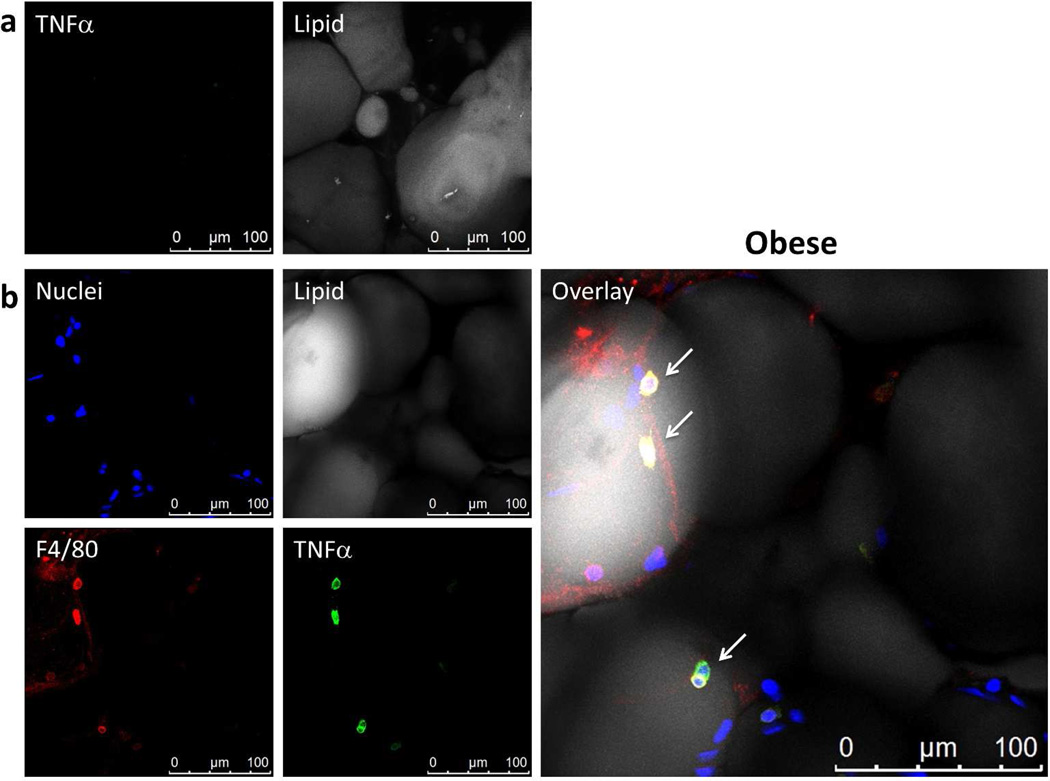
Adipocytes do not express TNFα. Whole adipose tissue was stained with BODIPY 558/568 for lipid (gray), Hoechst for nuclei (blue), anti-F4/80 for macrophages (red), anti-TNFα (green). All images were captured using LSCM with a 63× objective and are 2D projections of a 3D image stack. (a) Negative TNFα staining throughout the adipocytes visualized. Nuclei not shown. (b) TNFα staining was associated only with macrophages (arrows). No adipocytes stain for TNFα. Yellow in the overlay image indicates co-localization of TNFα and F4/80.

