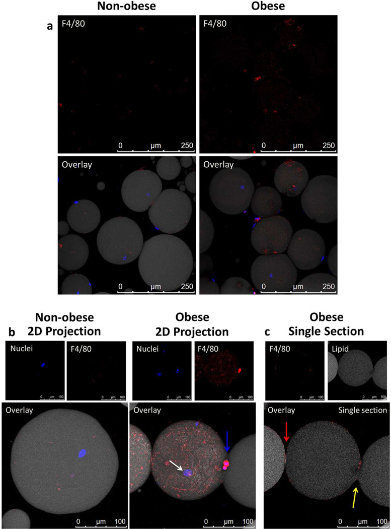
Figure 5.
Adipocytes in the floating layer were fully liberated from adipose tissue, yet macrophages remained bound to the single cell adipocytes, especially adipocytes recovered from obese mice. Floating layer cells were stained with anti-F4/80 (red) to stain macrophages, BODIPY 558/568 (grey) to stain lipid and Hoechst dye (blue) to stain nuclei. Anti-F4/80 primary antibody was visualized using anti-rat IgG Alexa647 conjugated secondary antibody. A sample from obese mice was also stained using the secondary antibody only and showed no background staining (data not shown) (a) Images show liberated, intact adipocytes after collagenase digestion and washing from non-obese and obese mice. The top panels show increased F4/80 covering adipocytes from obese mice when compared to non-obese mice. Images were collected with 20× objective. (b) Individual adipocyte images from obese and non-obese mice collected with 63× objective. In the obese condition, macrophages are seen covering individual intact adipocytes. An intact adipocyte was confirmed by the presence of an adipocyte nucleus (white arrow), while macrophage nuclei are morphologically different and stain more densely (blue arrow). (c) A one micron section taken from the middle of the 3D z-stack of the macrophage covered adipocyte in Figure 5b reveals that the large, single adipocyte is effectively being pinched in half by the macrophages surrounding it. This can be seen by the continuation of lipid (gray) between the attached circular bodies and the break in F4/80 staining on the right-hand side of the frame (yellow arrow) in contrast to the distinct border (red arrow) of macrophages covering the separate adipocytes on the left-hand side of the frame.

