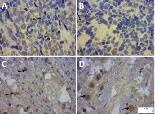
Figure 1.

Immunohistochemical staining of the biomarker QSOX1 in neuroblastoma slides (400 x). Note the positive staining in the cytoplasm, the extracellular matrix and, sometimes, the perinuclear space. The staining in the extracellular matrix is weaker in A) and B) and is more intense in C) and D). A) and B), poorly differentiated, Schwannianstroma poor neuroblastoma with unfavourable Shimada classification and low immunopositivity for QSOX1 (≤65 µm2). C) and D), well-differentiated, Schwannianstroma rich neuroblastoma with favourable Shimada classification and high immunopositivity for QSOX1 (≤65 µm2).
