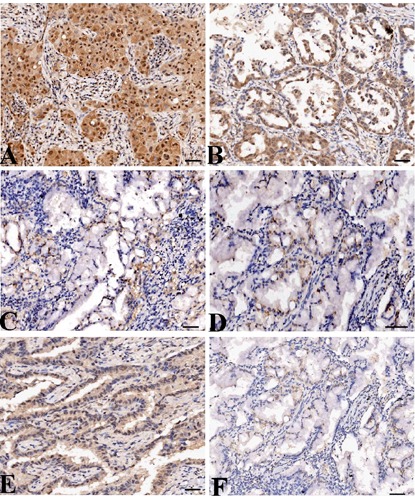
Figure 1.

The expression of MMP-7 protein in LAC tissues (magnification: 200×). LAC tissues were immunohistochemically stained with an anti-MMP-7 and COX-2 antibodies and classified as positive expression (A) and negative expression (C). Adjacent non-cancer tissues were immunohistochemically stained with an anti-MMP-7 antibody and classified as positive expression (B) and negative expression (D). COX-2 was highly expressed in LAC tissues (E) and lowly expressed in ANCT (F). Positive immunostaining of MMP-7 and COX-2 was mainly localized in the cytoplasm of tumor and tissue cells. Scale bars: A-C, E,F) 75 µm; D) 150 µm.
