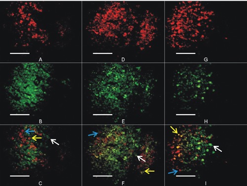
Figure 2.

Immunohistochemical images of buffalo fetus anterior pituitary gland during first, second and third trimester of gestation. (A; ACTH, B; POMC, C; co-localization of ACTH and POMC) at the age of 3 month fetus, (D; ACTH, E; POMC, F; co-localization of ACTH and POMC) at the age of 6 months, (G; ACTH, H; POMC, I; co-localization of ACTH and POMC) at 9 months of fetus age. Blue arrow shows individual cells having ACTH. White arrow shows individual cells having POMC granules, while yellow arrow shows the evidence of ACTH and POMC co-localization (yellow cells) in specific cell. Magnification: 400x.
