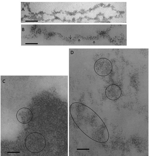
Figure 4.

A) Chromatin fibers spilling out of a ruptured chicken erythrocyte nucleus, negative staining with 0.5% ammonium molybdate adjusted to pH 7.4 to 8.0 with NH4OH. Scale bar: 50 nm. B,C,D) Thin sections from samples fixed in situ, resin embedded and stained with the Feulgen-like osmium-ammine reaction. B) regenerating rat hepatocytes, 24 h after partial hepatectomy; a long chromatin fiber is visible, 20-25 nm thick, which appears to be composed by a series of round particles with a diameter of about 11 nm, exhibiting an unstained inner core encircled by a DNA ring with a thickness of 2-3 nm (arrows). C) human resting lymphocyte; in the highly compact chromatin, particles similar to those described in (B) can be detected (encircled). D) regenerating rat hepatocyte; a particulate organization is present in the loosened chromatin structures (encircled). Scale bar: 50 nm
