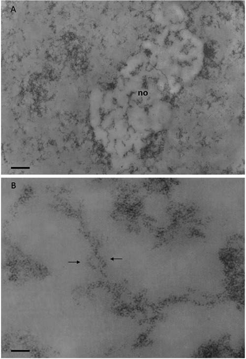
Figure 5.

Regenerating rat hepatocyte at 24 h after partial hepatectomy; Feulgen-like osmium-ammine staining. A) Chromatin is mainly in a dispersed form, only small clumps of compact chromatin can be detected; the nucleolar body (no) appears to be more electron-translucent than the remaining nucleoplasm, very likely due to RNA extraction; within the nucleolar body many chromatin fibers, well separated from each other, can be followed for a considerable length; scale bar: 0.4 μm. B) Loosened organization of the intranucleolar chromatin which appears to be composed of fibers with a thickness ranging from 11 to 20 nm; arrows indicate 11 nm thick chromatin fibers intertwined with each other, as the serpent around the rod of Asclepius; scale bar: 50 nm.
