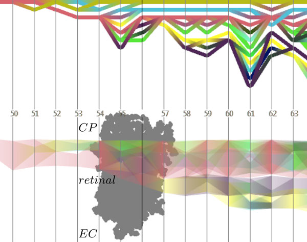
Figure 6.
Path timeline visualizations. The split and merge graph (top) shows topological events of the time-varying cavity structure. The evolution graph (bottom), on the other hand, depicts the position and size of the cavities along a user-defined direction. The time steps of the trajectory are given by the displayed numbers. At time step 55, a temporary connection between the cytoplasmic (CP) and the extracellular (EC) cavities occurs, which, however, does not represent the formation of a water-filled channel. To ease the understanding of the displayed events, a projection of the molecule is displayed in the evolution graph.

